
Patient Profile
Mr. Karthik S*********n, a 44-year-old construction supervisor from Pallikaranai, Chennai, was brought to the Emergency Department of a tertiary care trauma centre after a workplace accident at a high-rise building site in OMR. A heavy concrete slab had fallen onto his right foot, trapping it against a steel girder for nearly 20 minutes before co-workers could free him. He is the sole breadwinner for a family of four, and the fear of losing his foot — and his livelihood — was palpable from the moment he arrived at the Emergency Trauma Center.
Clinical Presentation
Mechanism of injury: Crush injury with high-energy blunt trauma
Time to presentation: 2.5 hours post-injury
Time to presentation: 2.5 hours post-injury
Examination Findings
-
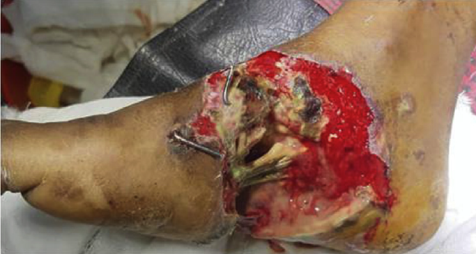
Right foot: Extensive degloving-type injury over the dorsum and medial aspect
-
Wound dimensions: 14 cm × 10 cm (approx. 140 cm²)
-
Wound bed: Heavily contaminated with construction debris (cement dust, rust particles)
-
Tissue viability: Extensive non-viable subcutaneous tissue; necrotic skin edges
-
Exposed structures: Extensor tendons to the second and third toes partially exposed; no gross bone involvement on initial probing
-
Neurovascular status: Dorsalis pedis pulse palpable but weak; sensation diminished over first web space
-
Radiographs: No fractures; soft tissue swelling significant
-
Infection markers: WBC 16.8 × 10⁹/L, CRP 210 mg/L
The injury was classified as a Gustilo-Anderson Type IIIB soft tissue injury by the orthoplastic team. Given the extensive contamination, tissue loss, and the high risk of progression to deep infection or necrosis, the team decided on immediate radical debridement followed by Vacuum-Assisted Closure (VAC) therapy as a bridging modality to prepare the wound for definitive soft tissue coverage.
Initial Surgical Management
Day 0: Emergency Debridement
- Under spinal anaesthesia in the operating theatre:
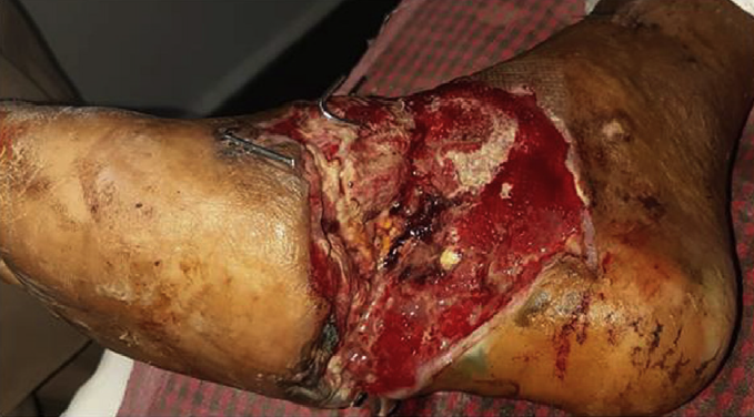
- All necrotic skin, subcutaneous fat, and devitalised fascia were radically excised until healthy, bleeding tissue was encountered.
- The wound was thoroughly irrigated with 10 litres of normal saline jet lavage to remove cement particulate matter.
- Meticulous haemostasis was achieved with bipolar cautery.
- The extensor tendons were preserved but left exposed with paratenon intact.
- Post-debridement, the defect measured 12 cm × 9 cm with significant dead space and a heavily exudative surface. Primary closure was impossible, and immediate flap coverage was deemed high-risk due to the contaminated field.
- The decision was made to apply NPWT to prepare the wound bed.
NPWT Protocol
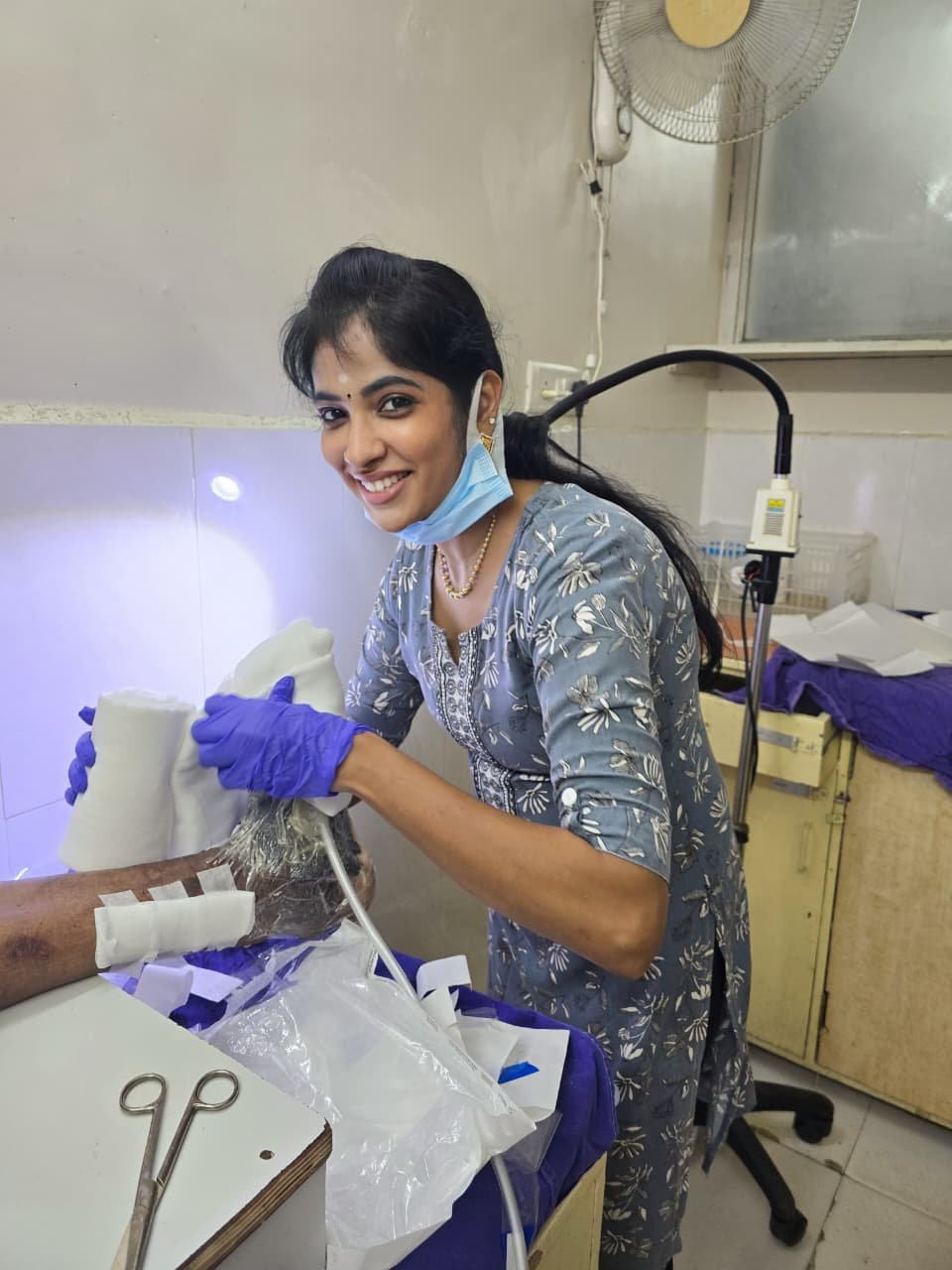
The institution utilised an indigenously assembled VAC system — a cost-effective approach described in Indian surgical literature — using locally available materials: Ortho cling drape, abdominal drain tubing, and sterile foam sponges, connected to wall-mounted suction. This brought the consumable cost down to approximately ₹2500 per dressing, compared to commercial systems that can exceed ₹10,000 per day — a critical consideration for a patient from a lower-middle-income household.
Foam Selection
Black polyurethane ether foam was selected for this large, exudative traumatic wound. As described in the literature, this foam is lighter, hydrophobic, and possesses an optimal pore size of 400–600 micrometres — ideal for distributing negative pressure evenly across an irregular wound bed and facilitating efficient fluid evacuation from a contaminated crush injury.
Pressure Settings & Mode : Negative pressure: −125 mmHg, Intermittent
-
Mode: Intermittent (5 minutes “on”, 2 minutes “off”)
-
Rationale: For acute traumatic wounds, −125 mmHg is considered the optimal pressure to stimulate granulation tissue formation without compromising perfusion. The intermittent mode was specifically chosen because studies demonstrate that the “suction off” phase generates a surge in blood flow through local vasodilation mediated by nitric oxide release — a phenomenon that does not occur with continuous suction. This cyclical hypoxia-reperfusion pattern stimulates angiogenesis and accelerates the proliferative phase of healing.
Dressing Change Schedule
The dressing was planned to be changed every 3 days (72 hours), with a commitment to complete at least 3 sessions before considering termination — as abrupt cessation after a single session can trigger a rebound phenomenon with worsening of the wound.
Mechanism of Healing: Why Intermittent NPWT Excelled
The dramatic improvement in Mr. Karthik’s crush injury can be attributed to the multimodal mechanisms of VAC therapy, as elucidated in contemporary reviews:
- Wound contraction and macro-strain: The −125 mmHg pressure generated mechanical forces that physically drew the wound margins inward, reducing the surface area requiring coverage.
- Microdeformation of cells: The foam’s 400–600 μm pores created microstrain across the wound bed, triggering cellular proliferation and growth factor release — essentially “exercising” the cells to divide and form granulation tissue.
- Oedema reduction and perfusion optimisation: Negative pressure removed interstitial fluid (following Bernoulli and Venturi principles), decompressing capillaries and improving oxygen delivery to the injured tissues.
- Angiogenesis via cyclical hypoxia: During the 2-minute “off” phase of intermittent therapy, tissue hypoxia beneath the foam stimulated a compensatory release of nitric oxide, causing vasodilation and angiogenic sprouting. This cyclical reperfusion is why intermittent mode achieves nearly twice the rate of granulation tissue formation compared to continuous mode.
- Bacterial clearance: The sealed environment, combined with active drainage of exudate (a bacterial growth medium), reduced the bioburden from a contaminated construction site wound to a sterile granulating bed within six days.
Key Learning Points
- Intermittent mode superiority: For acute traumatic wounds, the 5-minute-on, 2-minute-off cycle generates superior blood flow and angiogenesis compared to continuous suction. This should be the default setting in contaminated crush injuries with healthy vascular inflow.
- Optimal pressure: −125 mmHg is the evidence-based standard for acute traumatic wounds, balancing effective exudate removal and microdeformation against the risk of ischaemia.
- Foam selection matters: Black polyurethane ether foam (400–600 μm pore size) is ideal for large, exudative, contaminated wounds because its hydrophobic nature and lighter weight allow even pressure distribution and efficient fluid transport.
- Do not terminate abruptly: Completing a minimum of 3 sessions (with dressing changes every 3 days) prevents the rebound phenomenon and ensures durable granulation tissue formation.
Conclusion
Mr. Karthik’s case illustrates how a high-energy crush injury to the foot — traditionally a precursor to prolonged morbidity, deep infection, or amputation — can be transformed into a manageable wound through disciplined application of NPWT principles. By employing intermittent −125 mmHg suction with black polyurethane ether foam, changed every 3 days over three sessions, the team converted a contaminated, tendon-exposing wound into a graftable bed in just 9 days.
The mechanistic benefits — wound contraction, oedema reduction, microdeformation-driven cell proliferation, cyclical hypoxia-induced angiogenesis, and bacterial load clearance — worked synergistically to spare the patient from major flap reconstruction. For a construction worker in Chennai, this meant not just limb salvage, but the preservation of his family’s economic stability and his dignity as a provider.
Add a Comment