
Patient Profile
Mrs. Lakshmi Venkatesan, a 68-year-old retired schoolteacher from Chennai, Tamil Nadu, presented to the Department of Orthopaedic Surgery at a tertiary care centre in Chennai with a non-healing ulcer on the plantar surface of her left foot. A known case of Type 2 diabetes mellitus for 18 years with poor glycaemic control (HbA1c: 13.2%), she also had background diabetic neuropathy and hypertension. Despite six weeks of conventional saline gauze dressings and multiple courses of antibiotics prescribed at a local clinic, the ulcer had progressively enlarged, becoming heavily exudative with exposed plantar fascia — classified as a Wagner Grade 3 diabetic foot ulcer.
Her mobility had severely declined; she was largely bedbound and deeply distressed, having lost her independence and ability to perform daily prayers and visit the temple — activities central to her emotional well-being.
Clinical Presentation
On admission, examination revealed:
- Wound location: Plantar aspect of left foot, over the first metatarsal head.
- Wound dimensions: 8.4 cm × 6.2 cm (approx. 52 cm² surface area)
- Wound bed: Sloughy tissue with moderate seropurulent exudate
- Exposure: Plantar fascia visible; no bone involvement on probing
- Peripheral pulses: Diminished but palpable; Doppler showed monophasic flow
- Infection markers: Elevated WBC (14.2 × 10⁹/L), CRP (186 mg/L), ESR (118 mm/hr)
- Wound culture: Staphylococcus aureus and Bacteroides fragilis
Given the chronicity (42 days), failure of conventional dressings, and the patient’s deteriorating quality of life, the multidisciplinary diabetic foot team decided to initiate Vacuum-Assisted Closure (VAC) therapy following radical surgical debridement.
VAC Therapy Protocol
Initial Debridement
In the operating theatre, radical debridement was performed, excising all necrotic and infected tissue until healthy, bleeding granulation tissue was reached. The wound was thoroughly irrigated with jet lavage.
VAC Application
The wound was prepared using the standard VAC technique as described by Nather et al.
- A sterile polyurethane foam dressing was trimmed to fit the wound contours and placed into the defect.
- An adhesive drape was applied to cover the foam and an additional 3–5 cm of surrounding intact skin.
- A circular hole was created in the drape, and a non-collapsible tubing was secured over the opening.
- The tubing was connected to an electronic vacuum pump.
- Pressure Settings: A negative pressure of −125 mmHg was applied continuously, based on evidence that this setting achieves optimal macro-strain to draw wound edges inward and stimulate granulation tissue formation.
Course of Treatment & Dressing Changes
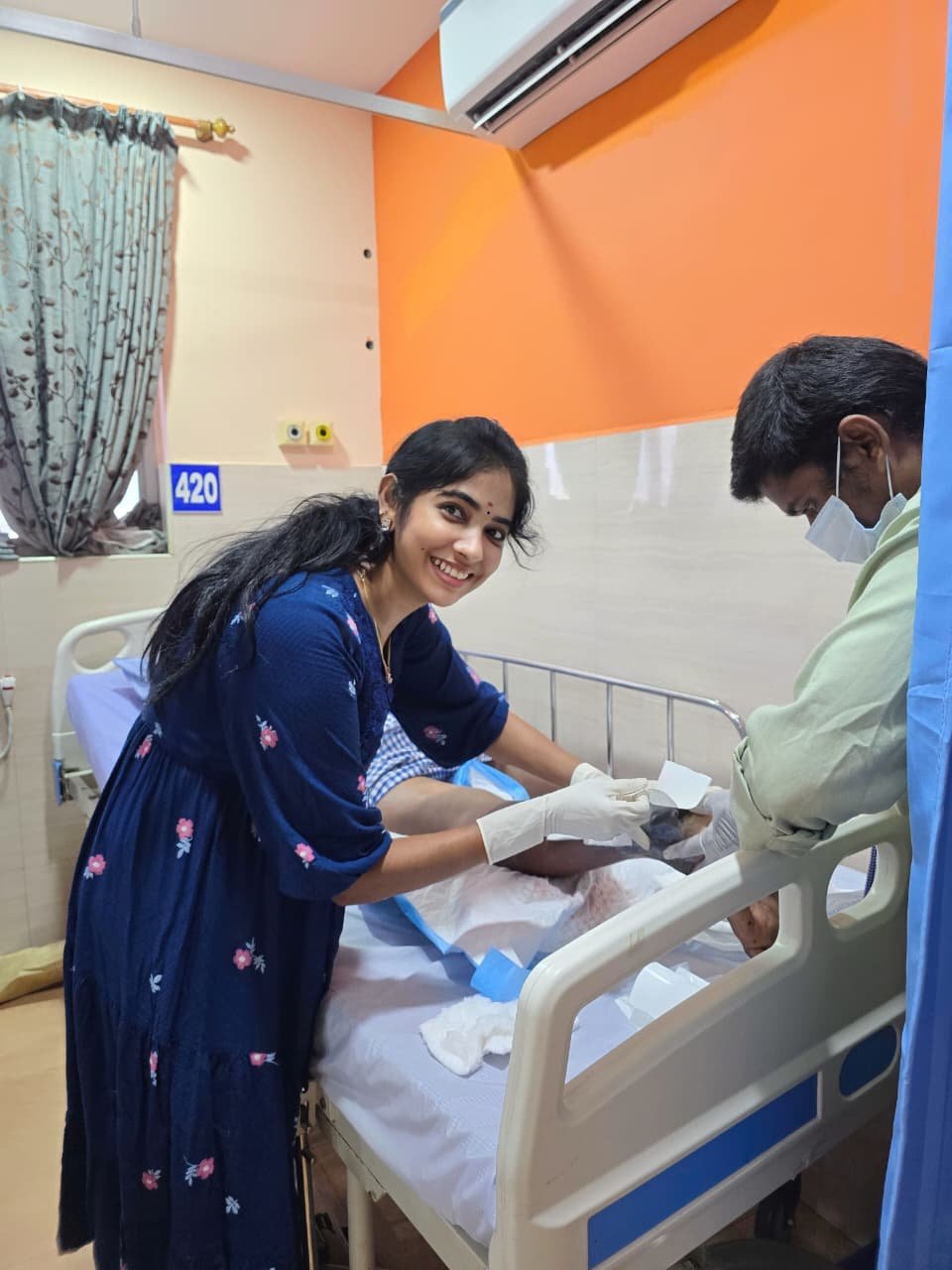
Mrs. Venkatesan’s VAC dressing was changed every 48 to 72 hours in the ward by a trained wound care nurse — a total of eight dressing changes over the course of her therapy.
At each dressing change:
- The wound was inspected for new slough or infection.
- The wound was cleansed with normal saline and hydrogen peroxide as indicated.
- Fresh foam and drape were reapplied.
- Wound dimensions were traced using the double polyethylene sheet technique over a 2 × 2 mm grid.
Surgical Closure & Final Outcome
On Day 19, Mrs. Venkatesan underwent split-thickness skin grafting in the operating theatre. The graft took completely with 100% viability by Day 26. She was discharged on Day 28 with instructions for off-loading footwear and strict glycaemic control.
Key Outcomes
- Total VAC therapy duration: 18 days
- Total dressing changes: 8
- Wound area reduction: 65.4%
- Infection status: Cleared by Day 12 (confirmed by negative wound culture)
- Final closure: Split-skin grafting with complete take
- Limb salvage: Successful — no amputation required
Impact on Patient Well-Being & Mobility
The most profound outcome was not merely the closure of the wound, but the dramatic transformation in Mrs. Venkatesan’s overall well-being.
For nearly two months, she had been confined to her home, dependent on her daughter for basic activities. The constant wound odour and fear of progression to amputation had left her anxious and withdrawn. Within days of VAC initiation, she reported significant pain relief — the occlusive, controlled environment of the VAC dressing eliminated the daily trauma of conventional gauze changes.
Once the ulcer healed and she was fitted with a diabetic footwear orthosis, Mrs. Venkatesan began walking independently within her apartment. By the sixth week post-discharge, she had resumed her morning walks to the neighbourhood Kapaleeshwarar Temple in Mylapore — a milestone she described as “getting my life back.” Her sleep improved, her appetite returned, and her HbA1c dropped to 7.8% over the next three months as she regained the confidence to manage her diabetes actively.
Her daughter noted: “Amma’s spirit returned the day she could walk to the temple again. The VAC dressing did not just heal her foot — it healed her mind.“
Add a Comment