
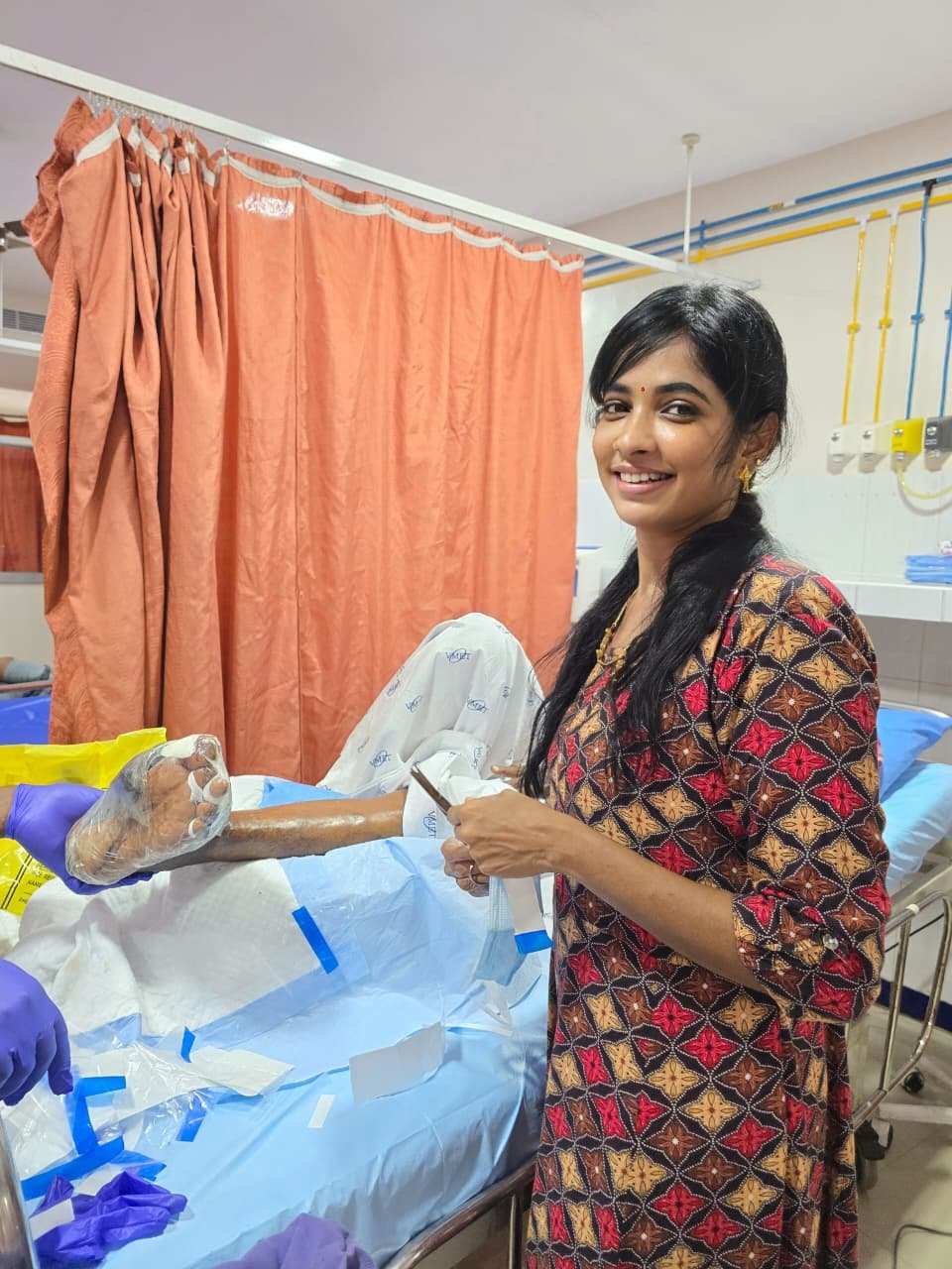
Patient Profile
Ms. Amritha R*******n, a 38-year-old home-maker residing in Adyar, Chennai, was admitted to the Department of Plastic and Reconstructive Surgery at a tertiary burn centre in Chennai following a domestic accident. While preparing a traditional Tamil feast for a family gathering, her silk saree caught fire from an open gas stove. Despite immediate attempts by family members to douse the flames, she sustained deep partial-thickness and full-thickness burns to both lower extremities.
A mother of two young children, Ms. Ramanathan was the primary caregiver in her joint family. The prospect of prolonged immobility and disfigurement caused her immense psychological distress from the very first day of admission.
Clinical Presentation & Initial Assessment
Mechanism of injury: Flame burns (domestic accident)
Time to presentation: 4 hours post-injury
- Right thigh (anteromedial) -> 8% TBSA (Deep partial to full-thickness)
- Left thigh (anterolateral) -> 7% TBSA (Deep partial to full-thickness)
- Right lower leg (anterior) -> 5% TBSA (Full-thickness )
- Left lower leg (anterior & medial) -> 6% TBSA (Deep partial to full-thickness)
- Total -> 26% TBSA
Clinical Findings on Day 3
-
Wound beds covered with dense, leathery eschar over the full-thickness areas
-
Underlying slough in deep partial-thickness zones
-
Moderate serosanguinous exudate
-
No signs of inhalation injury
-
Vitals stable; haemoglobin 10.2 g/dL
-
Pain score: 8/10 despite analgesia
After resuscitation and stabilisation, the surgical team decided that the full-thickness and deep dermal components would not heal spontaneously within a reasonable timeframe and would result in severe hypertrophic scarring and contractures if left to secondary intention. The plan was early tangential excision and split-thickness skin grafting (STSG) — the gold standard for deep burns of this extent.
Surgical Management : Day 4: Tangential Excision
Under general anaesthesia, the burn wounds were subjected to tangential excision using a Watson knife. The eschar was sequentially shaved until viable, bleeding dermis and subcutaneous tissue were encountered. The excised areas included:
-
Right thigh: 18 cm × 14 cm defect (approx. 252 cm²)
-
Left thigh: 16 cm × 12 cm defect (approx. 192 cm²)
-
Right lower leg: 14 cm × 10 cm defect (approx. 140 cm²)
-
Left lower leg: 15 cm × 11 cm defect (approx. 165 cm²)
Total excised area: ~749 cm²
The wounds were copiously irrigated with warm saline. Haemostasis was meticulously achieved using adrenaline-soaked gauze and pinpoint electrocautery.
Day 4: Split-Thickness Skin Grafting
A 0.012-inch (0.30 mm) split-thickness skin graft was harvested from the left anterolateral thigh using a pneumatic dermatome. The graft was meshed at a 1.5:1 ratio to allow expansion and drainage, then applied to all four excised sites.
NPWT Application for Graft Bolstering
Traditionally, the team would have secured these grafts with tie-over bolster dressings or elastic crepe bandages. However, given the large surface area, the mobile nature of the thigh tissue, and the risk of shear forces during patient movement, the senior plastic surgeon elected to use Vacuum-Assisted Closure (VAC) therapy as a graft bolstering system.
Method of Application
Following the graft placement:
- Non-adherent interface: A single layer of Vaseline-impregnated gauze was placed loosely over the meshed graft to prevent the foam from adhering to the delicate neo-epidermis.
- Polyurethane foam: Sterile black VAC foam was cut to size and placed over each grafted site. Care was taken to ensure the foam extended 2 cm beyond the graft margins.
- Adhesive drape: Transparent adhesive film was applied to create an airtight seal over the foam and surrounding intact skin (3–5 cm margin).
- Tubing & suction: The suction pad was placed centrally, and the tubing was connected to a portable electronic vacuum pump.
Pressure Settings: Negative pressure: −75 mmHg applied continuously
Rationale: A lower pressure of −75 mmHg was chosen (rather than −125 mmHg) because:
- The wounds were fresh grafts, not chronic ulcers.
- Higher pressures can theoretically compromise graft viability in the first 48–72 hours.
- Evidence from plastic surgery literature supports −50 to −75 mmHg for graft fixation to optimise take without inducing shear stress on the immature graft.
Total VAC therapy duration: 12 days
Total dressing changes: 4 (at 72-hour intervals)
Average drainage per change: 30–50 mL of serous fluid
Clinical Outcomes
Graft Survival
-
Overall graft take: 97.3% — comparable to published series reporting ~96% take with NPWT bolstering
-
No haematoma or seroma formation requiring evacuation
-
No evidence of graft displacement or shear injury despite the patient being allowed gentle knee flexion from Day 5
Infection & Complications
-
Wound cultures at Day 3 and Day 9: negative for bacterial growth
-
No systemic signs of infection (afebrile throughout)
-
No maceration of surrounding skin
Pain & Patient Comfort
Ms. Ramanathan reported that the VAC system was far more comfortable than the tight elastic bandages she had anticipated. The continuous suction eliminated the “wet, heavy” feeling of traditional dressings. Her pain score dropped from 8/10 pre-operatively to 3/10 by Day 3, and she required only oral paracetamol and tramadol by Day 5.
Rehabilitation & Return to Mobility
The psychological impact of the burn had initially left Ms. Ramanathan withdrawn and fearful. However, the reliability of the VAC dressing allowed the physiotherapy team to begin early, aggressive rehabilitation:
-
Day 3: Passive range-of-motion exercises for knees and ankles while in bed
-
Day 7: Assisted standing with walker (VAC tubing managed with a portable holster)
-
Day 10: Independent ambulation within the ward
-
Day 14: Discharged home with compression garments and silicone gel sheets
By Week 6, she was walking unaided to her children’s school in Adyar. The grafted areas healed with minimal hypertrophic scarring compared to historical controls in the same unit, which the surgical team attributed to the uniform pressure distribution and reduced inflammatory milieu created by NPWT.
At her 6-month follow-up,
Ms. Ramanathan expressed profound gratitude: “I was terrified I would never wear a saree again or bend down to pick up my daughter. The VAC dressing held my new skin in place so perfectly that I healed completely with no visibly raised scars. I got my life back — and my confidence.“
Add a Comment